Lincolnshire Knee Clinic
Treatments

Realignment Procedures (Osteotomy)

Bone-cutting procedures to correct knee alignment and shift load away from a damaged compartment, delaying or avoiding joint replacement.

On this page

Overview

Osteotomy (from the Greek for 'bone cutting') is a precision surgical procedure that corrects the mechanical axis of the leg. By carefully realigning the bones around the knee, the procedure offloads a damaged or arthritic compartment and transfers stress onto healthier cartilage. This page covers high tibial osteotomy (HTO), distal femoral osteotomy (DFO), and slope-changing osteotomy.

An osteotomy involves making a precise angular cut through bone — either the upper tibia (shinbone) or lower femur (thighbone) — to correct the mechanical alignment of the leg. Rather than replacing the joint surface, the procedure redistributes the load across the knee so that a healthy compartment bears more of the body's weight, relieving pressure from the damaged side. The corrected position is held by a steel plate and screws. There are three main variants used at this clinic: High Tibial Osteotomy (HTO): The most common type. A wedge is opened or closed in the upper tibia to correct a bow-legged (varus) deformity, shifting load away from the worn medial (inner) compartment onto the healthy lateral (outer) side. Particularly suited to active, younger patients with medial knee arthritis or post-ligament-surgery malalignment. Distal Femoral Osteotomy (DFO): A cut through the lower femur to correct a knock-kneed (valgus) deformity, offloading the worn lateral compartment. This is indicated when the valgus deformity originates in the femur rather than the tibia. Slope-Changing Osteotomy: A modification of HTO or DFO that also changes the posterior slope of the tibial plateau (the angle of the top of the tibia). Reducing tibial slope is used to protect ACL grafts or treat specific ACL graft failure patterns by reducing anterior tibial translation forces.
This information is for general patient education and does not replace an individual clinical assessment. Suitability and outcomes depend on joint condition and physical health.

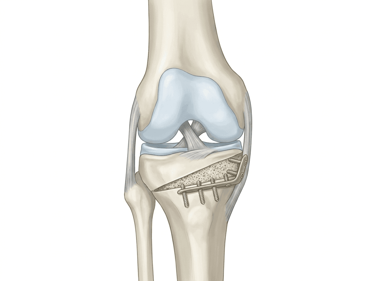
Clinical Overview Illustration

Medical illustration of a high tibial osteotomy showing an angular bone wedge opened in the upper tibia and secured with a titanium locking plate and screws to correct varus knee alignment.
Opening-wedge high tibial osteotomy: the corrected mechanical axis is maintained by a titanium locking plate while the bone gap heals..

What does this treatment involve?

Typical clinical stages of this treatment pathway include:

1Comprehensive pre-operative planning using full-leg weight-bearing X-rays to measure mechanical axis deviation and calculate the precise correction angle
2Spinal or general anaesthetic and surgical preparation; tourniquet applied to the thigh
3For HTO: a small incision below the kneecap on the inner side of the tibia; the bone is precisely cut and the wedge is opened to the calculated angle using spreader pins under X-ray guidance
4For DFO: an incision on the outer lower thigh; a closing or opening wedge cut is made in the distal femur to correct valgus deformity
5For slope-changing osteotomy: the tibial cut is angled to simultaneously alter the posterior tibial slope, reducing the resting forward pull on the ACL
6Rigid fixation of the corrected position with a low-profile titanium locking plate and screws
7The bone gap (in opening wedge osteotomies) may be filled with bone graft or a synthetic bone substitute
8Wound closure, compression dressings, and a protective knee splint or brace

Suitability

Suitability depends entirely on an individual clinical examination, diagnostic imaging, medical history, and personal activity goals.

Who may benefit?

  • Active patients under 60 (though age alone is not an absolute contraindication) with unicompartmental knee arthritis or early cartilage damage
  • Patients with medial compartment arthritis and a varus (bow-legged) deformity — candidates for HTO
  • Patients with lateral compartment arthritis and a valgus (knock-kneed) deformity — candidates for DFO
  • Patients with ACL graft failure or recurrent instability associated with an elevated posterior tibial slope — candidates for slope-changing osteotomy
  • Patients wishing to remain physically active and defer or avoid knee replacement
  • Patients with isolated compartment wear and healthy ligaments

When might it not be suitable?

  • Patients with severe, widespread (tricompartmental) osteoarthritis affecting all three compartments
  • Inflammatory arthritis (e.g. rheumatoid arthritis) causing global joint destruction
  • Severe joint stiffness with reduced range of movement before surgery
  • Patients unable to comply with the post-operative non-weight-bearing or restricted weight-bearing period
  • Significant bone loss or poor bone quality that would not hold the fixation plate securely
  • Active smokers (impairs bone healing significantly)

Alternatives

Alternative treatment pathways that may be considered depending on clinical severity:

Conservative management: physiotherapy, weight management, and unloading knee bracesExplore Option >
Clinical joint injections (steroid, hyaluronic acid, or biological agents) for pain managementExplore Option >
Partial knee replacement (unicompartmental), if the patient is older or less activeExplore Option >
Total knee replacement (for more advanced, multi-compartment disease)Explore Option >

Risks

As with any clinical intervention, risks and complications differ between patients. Suitability and risk profiles are discussed in detail during consultation. General risks associated with this procedure include:

Under- or over-correction of the alignment — may require revision or alter the plan for future joint replacement
Non-union or delayed healing of the osteotomy site (risk increased by smoking)
Hardware irritation from the plate and screws — occasionally requires removal once healed (typically 12–18 months post-op)
Deep vein thrombosis (DVT) or pulmonary embolism — blood thinners are used to reduce this risk
Infection at the surgical site or around the metalwork
Peroneal nerve stretch injury (particularly DFO or lateral HTO), causing temporary foot weakness or numbness
Fracture propagation beyond the planned cut if bone quality is poor
Progression of arthritis in the offloaded or remaining compartments over time

Recovery & Rehabilitation

Post-treatment rehabilitation and timelines are key to restoring joint health. We do not provide guaranteed recovery times.

Immediate Recovery

  • Weight-bearing is typically restricted for 6 weeks after opening-wedge osteotomy while the bone gap heals — crutches are required
  • Radiographic bone healing is confirmed by X-ray at 6–8 weeks before weight-bearing is progressed
  • Driving is typically possible at 6–8 weeks once crutch-free and safe muscle control is confirmed
  • Return to desk work is often possible at 6–8 weeks if crutch-independent
  • Return to manual work or sport takes 4–6 months; full recovery and confidence in the knee continues to improve for 12 months
  • The hardware plate is usually retained permanently unless symptomatic

Rehabilitation Pathway

  • Immediately post-operatively: quad activation, ankle pumps, and protected range of motion exercises in the brace
  • Weeks 1–6: restricted weight-bearing on crutches; daily physiotherapy for swelling control, range of motion, and isometric strengthening
  • Weeks 6–12: progressive weight-bearing once healing confirmed; focus on gait retraining and quadriceps strengthening
  • Months 3–6: functional strengthening, proprioception training, and progressive return to low-impact activity
  • Months 6–12: gradual return to sport-specific loading and higher-impact activities as directed by the surgeon
Rehabilitation Milestones

Realignment Procedures (Osteotomy) Recovery Timeline

Typical phase-based progression and return to functional activity guidelines.

1
Phase 1 (Early Stage)
Immediate Recovery & Adaptation
Primary Focus:Pain control, local tissue protection, and load modification.
  • Weight-bearing is typically restricted for 6 weeks after opening-wedge osteotomy while the bone gap heals — crutches are required
  • Radiographic bone healing is confirmed by X-ray at 6–8 weeks before weight-bearing is progressed
  • Driving is typically possible at 6–8 weeks once crutch-free and safe muscle control is confirmed
  • Return to desk work is often possible at 6–8 weeks if crutch-independent
  • Return to manual work or sport takes 4–6 months; full recovery and confidence in the knee continues to improve for 12 months
  • The hardware plate is usually retained permanently unless symptomatic
2
Phase 2 (Active Rehab)
Strength & Movement Restoration
Primary Focus:Targeted exercise to rebuild joint capacity and confidence.
  • Immediately post-operatively: quad activation, ankle pumps, and protected range of motion exercises in the brace
  • Weeks 1–6: restricted weight-bearing on crutches; daily physiotherapy for swelling control, range of motion, and isometric strengthening
  • Weeks 6–12: progressive weight-bearing once healing confirmed; focus on gait retraining and quadriceps strengthening
  • Months 3–6: functional strengthening, proprioception training, and progressive return to low-impact activity
  • Months 6–12: gradual return to sport-specific loading and higher-impact activities as directed by the surgeon
Important Clinical AdviceRecovery timelines are indicative. Individual rehabilitation progression depends on surgical findings, tissue quality, and the specific guidance of your consultant orthopaedic surgeon and physiotherapist.

Surgical Recovery Resources

As this is a surgical procedure, you may find our recovery resources helpful. These explain how to prepare and what to expect:

Clinical Pathway Map

Staged treatment and recovery journey.

Anatomy Comparison

Management Strategy Comparison

Comparing conservative treatment with active intervention options.
Conservative Path
Conservative PathConservative PathClinical Reference
Interventional Path (Intervention Options)
Interventional PathInterventional PathClinical Reference
Primary Focus

Conservative Path

Symptom management, quad strength, offloading

Interventional Path

Structural correction, joint resurfacing or repair

Timeframe

Conservative Path

Gradual ongoing adaptation over 3-6 months

Interventional Path

Structured acute healing followed by targeted rehab

Expected Outcome

Conservative Path

Improved function and pain control, preserved anatomy

Interventional Path

Restored mechanical stability, reconstructed tissue

Frequently Asked Questions

Both procedures realign the leg, but they target different bones. HTO corrects bow-leggedness (varus) originating in the tibia and is used for medial compartment arthritis. DFO corrects knock-knee (valgus) originating in the femur and is used for lateral compartment arthritis. The choice depends on where the deformity originates — determined by long-leg X-ray measurements.
Yes. In appropriately selected younger, active patients, a well-performed osteotomy can delay the need for knee replacement by 10 to 15 years or more. It preserves the native knee joint and all its ligaments, which is particularly important for patients who wish to stay physically active.
A slope-changing osteotomy modifies the backwards tilt (posterior slope) of the top of the tibia during an HTO. Reducing this slope decreases the forward pull on the ACL, and is used specifically to protect a reconstructed ACL or to address recurrent ACL graft failures related to an excessively steep tibial slope.
Not necessarily. Many patients retain the hardware indefinitely without any problems. However, if the plate causes local irritation or discomfort, it can be removed once the bone has fully healed — usually 12 to 18 months after surgery.
The bone cut typically shows X-ray evidence of bridging callus (early healing) by 6–8 weeks. Complete bone consolidation takes approximately 3–4 months, after which full weight-bearing and progressive activity are allowed.
Awaiting Clinical Review

This page is in draft and has not yet been formally reviewed by our clinical team. Content may change following clinical review.

References
  1. British Orthopaedic Association — High Tibial Osteotomy
  2. NICE — Osteotomy for knee osteoarthritis
  3. Lobenhoffer P, Agneskirchner JD. Improvements in surgical technique of valgus high tibial osteotomy. Knee Surg Sports Traumatol Arthrosc. 2003;11(3):132–8.

Would you like to discuss your treatment options?

A consultation can help clarify the cause of your symptoms and discuss appropriate treatment options.

Face-to-face appointments are arranged through the reception team at the chosen clinic. Video consultations use Google Calendar where available.

Continue Your Knee Journey

Next Step: Recovery & Rehabilitation

Understand recovery milestones, physical therapy timelines, and guidelines for returning to daily activities, driving, work, or sports safely.

Explore Recovery

Get notified about new content on Realignment Procedures (Osteotomy)

We'll only email you about updates on this specific topic — unsubscribe anytime.

Medical Disclaimer

This website provides educational and general information about knee symptoms, conditions, and treatments. The content is for informational purposes only and does not constitute medical advice, diagnosis, or a treatment plan. It should not be used as a substitute for a professional consultation, examination, or clinical decision-making by a qualified orthopaedic specialist.

If you are experiencing symptoms or are concerned about a knee problem, please arrange a clinical consultation. If you have urgent symptoms, severe pain, or are unable to put weight on your leg, please read our urgent advice guidance or contact emergency services immediately (dial 999 or NHS 111).

Contact us on WhatsApp — for administrative enquiries only. Please do not send confidential medical information.