Lincolnshire Knee Clinic
Conditions

Knee Arthritis

Knee arthritis is a common cause of pain, stiffness and reduced mobility. This page explains how it may affect the knee, how it is assessed and the treatment options that may be considered.

On this page

What is Knee Arthritis?

Knee arthritis is a clinical condition characterised by the gradual loss or damage of the smooth articular cartilage lining the joint surfaces. As the cartilage thins and wears, the underlying bone surfaces can experience friction, narrowing the joint space. This can lead to joint stiffness, pain, and reduced function. Wording is cautious: symptoms vary greatly between individuals, and X-ray findings do not always correlate with symptom severity.
This information is for general patient education and does not replace an individual clinical assessment.

Anatomical Overview

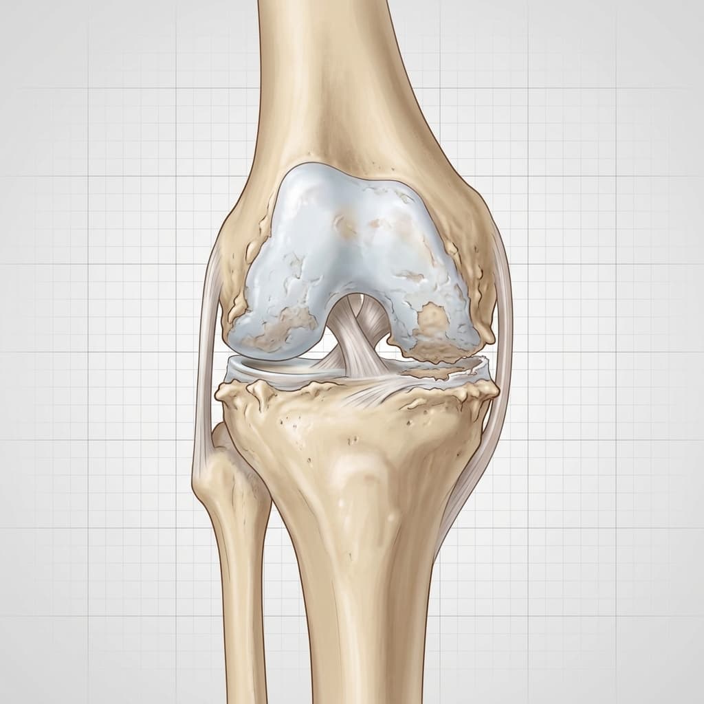
Diagram of a knee joint with osteoarthritis, highlighting loss of articular cartilage, joint space narrowing, and bone spurs.
Cartilage Degeneration

Thinning and erosion of the protective articular cartilage.

Osteophytes (Bone Spurs)

Bony projections forming along the joint margins in response to load changes.

Narrowed Joint Space

Reduced clearance between femur and tibia due to cartilage loss.

Anatomical changes associated with osteoarthritis of the knee, showing cartridge degradation.. (Pulsing letters show planned interactive annotation markers)
Cartilage DegenerationPathology

Thinning and erosion of the protective articular cartilage.

Osteophytes (Bone Spurs)Pathology

Bony projections forming along the joint margins in response to load changes.

Narrowed Joint SpacePathology

Reduced clearance between femur and tibia due to cartilage loss.

Anatomy Comparison

Healthy vs Affected Joint Comparison

Understanding structural differences caused by knee arthritis.
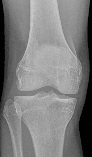
Normal Anatomy
Normal AnatomyNormal X-rayClinical Reference
Pathological / Affected Anatomy (Clinical Highlight)
Pathological / Affected AnatomyArthritic X-rayClinical Reference
Joint Space

Normal Anatomy

Wide, even clearance between bone surfaces

Pathological / Affected Anatomy

Narrowed joint space due to tissue degradation

Structural Integrity

Normal Anatomy

Smooth articular cartilage and strong stabilizing bands

Pathological / Affected Anatomy

Eroded surfaces, osteophytes (bone spurs), or exposed subchondral bone

Interactive Knee Anatomy

Educational Anatomy Illustration

Interactive Knee Anatomy (Textbook Engraving) — Osteoarthritis

Select any numbered hotspot on the anatomy book illustration below to view details. Use the category filters to simplify the view.
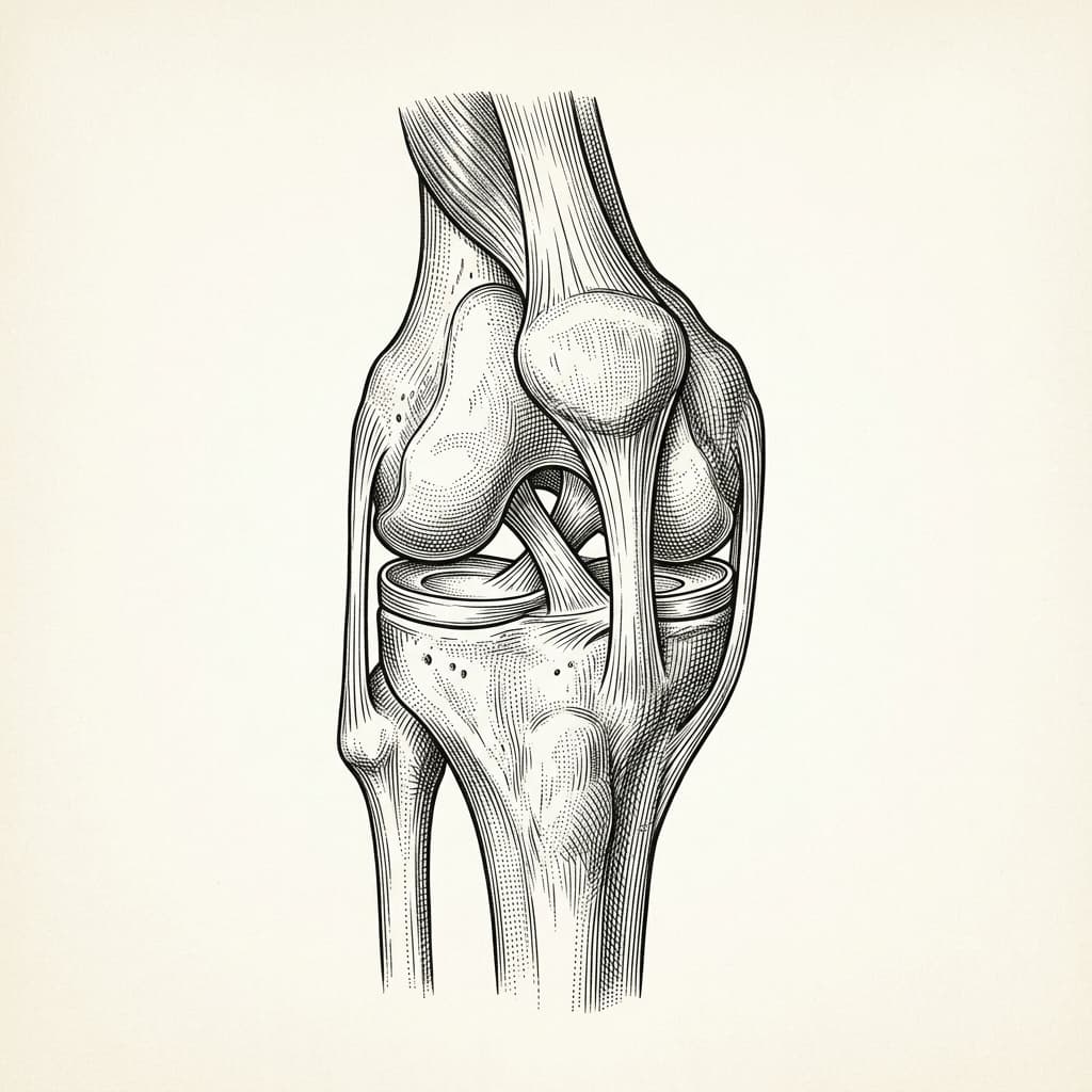
Educational illustration — awaiting clinical review before use as a clinical reference.
Anatomy book engraving of the human knee joint
Anatomy Details by Structure
View all structures as text list
  • Femur (Thigh Bone)

    The lower end of the femur forms the upper part of the knee joint. The rounded medial and lateral condyles glide and pivot on the tibial plateau.

  • Patella (Kneecap)

    A triangular sesamoid bone embedded in the quadriceps and patellar tendon complex. It glides through the trochlear groove, protecting the joint front and multiplying quadriceps leverage.

  • Tibia (Shin Bone)

    The main weight-bearing shin bone. The flat tibial plateau forms the lower surface of the knee joint, carrying the menisci.

  • Fibula (Calf Bone)

    The smaller outer shin bone. It does not carry joint weight directly but serves as an attachment point for the lateral collateral ligament (LCL) and biceps femoris muscle.

  • Articular Cartilage

    Smooth, wear-resistant hyaline cartilage that caps the ends of the femur and tibia. It provides an extremely low-friction surface, distributing weight and protecting subchondral bone.

  • Medial Meniscus

    A thick, C-shaped fibrocartilage shock absorber on the inner side of the knee joint. It spreads load, improves bone congruency, and aids joint stability.

  • Lateral Meniscus

    A circular fibrocartilage shock absorber on the outer side. It is slightly more mobile than the medial meniscus, assisting load transmission across the lateral joint compartment.

  • Anterior Cruciate Ligament (ACL)

    A key stabilizing ligament running diagonally deep inside the joint. It prevents the tibia from sliding forward on the femur and controls knee pivoting rotation.

  • Posterior Cruciate Ligament (PCL)

    The strongest ligament inside the knee, crossing behind the ACL. It prevents the tibia from sliding backward on the femur.

  • Patellar Tendon

    A strong fibrous band connecting the patella bottom to the tibial tuberosity on the shin bone, transmitting quadriceps extension force.

  • Quadriceps Tendon

    The thick tendon connecting the quadriceps muscles to the top of the patella. Essential for extending the knee joint.
Educational illustration only. This diagram is an educational representation of knee anatomy and is awaiting formal clinical review. It is not a clinical reference, does not represent any specific patient, and must not be used for diagnosis or treatment decisions.

Annotated Imaging Example

LKC Imaging Viewer
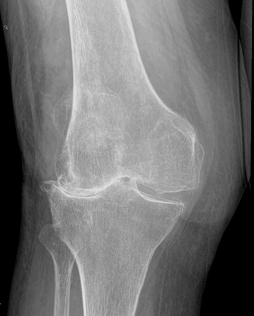
X-ray · AP (anterior-posterior) weight-bearing
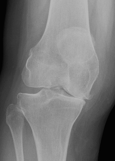
Weight-bearing AP X-ray of an arthritic knee showing narrowing of the medial joint space and increased bone density beneath the worn cartilage zone. Annotation markers highlight key radiological changes.
Annotations visibleEducational use only
Weight-bearing AP X-ray demonstrating medial compartment osteoarthritis. Educational example only.
Annotation Reference
  1. 1Medial Joint Space Narrowing
  2. 2Subchondral Sclerosis
  3. 3Lateral Compartment (Preserved)
Educational Imaging DisclaimerThis imaging example is provided solely for educational purposes. It is not a diagnostic image and does not represent your own anatomy or pathology. All patients require individual clinical examination, history taking, and appropriate investigations to reach a diagnosis. The findings shown may not be present in your case.

Common symptoms

Symptoms vary, and similar symptoms can occur with other knee conditions. Commonly reported signs include:

Knee pain (especially during weight-bearing or walking)
Joint stiffness, particularly in the morning
Reduced movement and difficulty bending the knee
Intermittent swelling or joint warmth
Crepitus (grinding or creaking sensations) during motion

Causes and associated factors

The development or onset of this condition can be associated with several mechanical, biological, and lifestyle factors, including:

  • Age-related degenerative wear of joint surfaces
  • Previous knee trauma or fractures (post-traumatic arthritis)
  • Previous meniscal or ligament injury altering loading
  • Leg alignment factors or genetic predispositions

Clinical assessment and diagnosis

A comprehensive clinical assessment is performed to understand the root cause of your symptoms and outline appropriate management options.

  • Discussion of symptoms, duration, and mechanical triggers
  • Assessment of impact on work, sleep, walking, and daily activity
  • Review of previous knee injuries, operations, and treatments
  • Physical examination of alignment, movement, swelling, and stability
  • Evaluation of patient goals and review of existing imaging
A diagnosis should not be made from website information alone.

Investigations

Imaging and other investigations may be recommended depending on clinical presentation. Common assessments include:

Weight-bearing X-rays to assess joint space narrowing and bone spurs

Indicated where clinically appropriate following assessment.

Diagnostics & Imaging >

MRI scans

if diagnostic uncertainty or concurrent soft-tissue pathology exists

Diagnostics & Imaging >

Blood tests

to rule out inflammatory arthritis in selected cases

Diagnostics & Imaging >
LKC Imaging Console v1.0
Status: clinically approved
Weight-bearing X-ray of an osteoarthritic knee demonstrating asymmetrical medial joint space narrowing.
Medial Joint Space Narrowing

Indicates loss of cartilage loading surface on the inner side of the knee.

Subchondral Sclerosis

Increased bone density (whitening) under the worn cartilage zone.

Weight-bearing anterior-posterior (AP) X-ray showing medial joint space narrowing.. (Weight-bearing views are standard diagnostic protocols)
Medial Joint Space NarrowingPathology

Indicates loss of cartilage loading surface on the inner side of the knee.

Subchondral SclerosisPathology

Increased bone density (whitening) under the worn cartilage zone.

Treatment options

We offer structured, staged treatment pathways, focusing on conservative and non-surgical approaches. Suitability depends on individual assessment.

A

Education and lifestyle or activity modification

Indicated where clinically appropriate.

Explore Treatment >
B

Physiotherapy and quadriceps strengthening exercises

Indicated where clinically appropriate.

Explore Treatment >
C

Weight management strategies to reduce mechanical load

Indicated where clinically appropriate.

Explore Treatment >
D

Pain-relieving medication and clinical joint injections

Indicated where clinically appropriate.

Explore Treatment >
E

Surgical replacement

partial or total knee replacement) when appropriat

Explore Treatment >

When might surgery be considered?

Surgical intervention may be discussed when non-operative treatments do not provide sufficient relief, symptoms significantly restrict daily function or quality of life, and clinical and imaging findings correlate.

Partial Knee Replacement

unicompartmental) for single-compartment wea

Total Knee Replacement for widespread tricompartmental degeneration

Consultant orthopaedic procedure.

Choosing the right treatment

Decisions depend on a detailed diagnosis, symptom severity, patient goals, activity expectations, and general health history. We support shared decision-making, ensuring you fully understand the expected benefits, risks, and alternatives of each pathway.

Frequently Asked Questions

No, surgical intervention is not always required. We prioritise non-surgical joint preservation strategies, such as physical therapy, activity modifications, and clinical knee injections, recommending surgery only when conservative options have been fully explored and joint pain continues to restrict your quality of life.
Hospital stay is usually 1-3 days. Patients typically walk with aids for 4-6 weeks and require structured physiotherapy. Significant pain relief and functional improvement are observed by 3 months, with full recovery continuing up to 1 year.
Knee arthritis is typically a progressive condition, meaning it can change over time. However, the rate of progression varies significantly between individuals. Many patients manage symptoms effectively for years using non-surgical strategies like physical therapy, weight management, and targeted injections, without requiring surgery.
No, an MRI is not required for every patient. In most cases of knee arthritis, a detailed clinical examination and standard weight-bearing X-rays are sufficient to establish a diagnosis. An MRI may be recommended if there is diagnostic uncertainty, or if another soft-tissue condition (such as a meniscus tear or ligament injury) is suspected.
No, appropriate low-impact exercise does not make knee arthritis worse. In fact, exercise is a primary recommendation for managing symptoms. Strengthening the muscles surrounding the joint (such as the quadriceps) and maintaining flexibility can reduce pain and improve knee stability. High-impact or repetitive overloading, however, should be managed carefully.
No, knee injections are not suitable for all individuals. Suitability depends on the severity of the arthritis, your medical history, and any existing health conditions (e.g., active infections or uncontrolled diabetes). Benefits vary and are often temporary. A full clinical discussion is required to evaluate the risks and alternatives.
Knee replacement surgery is typically discussed when knee pain and stiffness significantly restrict your daily activities, sleep, or mobility, and when appropriate non-surgical treatments (such as physical therapy and injections) have failed to provide sufficient relief. The decision is made through a shared process between you and your consultant.
Yes, maintaining active participation in work and low-impact exercise is generally encouraged. It helps preserve joint range of motion and overall health. Modifying activities—such as swimming or cycling instead of high-impact running—and taking regular breaks can help manage symptoms while keeping you active.
Yes. The knee joint has three main compartments: medial (inside), lateral (outside), and patellofemoral (under the kneecap). Arthritis can be localized to just one compartment (unicompartmental arthritis) or affect multiple areas (tricompartmental arthritis). If localized, a partial knee replacement may be a clinical option.
While a GP referral is highly recommended and often required by private medical insurance companies to authorize cover, self-paying patients can typically self-refer and schedule a consultation directly with the clinic. We recommend checking with your insurance provider prior to booking.
Clinically Reviewed
Mr Ricardo J Pacheco (GMC 4145976)Consultant Trauma & Orthopaedic Surgeon
Last reviewed: 29 July 2026
References
  1. NICE guideline NG226 — Osteoarthritis in over 16s: diagnosis and management
  2. NHS — Osteoarthritis
  3. Osteoarthritis in over 16s: diagnosis and management | Guidance (nice.org.uk)
  4. Validation of NICE Criteria for the Diagnosis of Knee Osteoarthritis (acrabstracts.org)
  5. Evidence Sources: Osteoarthritis of the Knee Clinical Care Standard (safetyandquality.gov.au)
  6. Management of Osteoarthritis of the Knee (Non-Arthroplasty) (aaos.org)

Would you like an individual knee assessment?

A consultation can help clarify the cause of your symptoms and discuss appropriate treatment options.

Face-to-face appointments are arranged through the reception team at the chosen clinic. Video consultations use Google Calendar where available.

Continue Your Knee Journey

Next Step: Explore Treatment Options

Treatment pathways range from physiotherapy and bracing to injections or surgical procedures, tailored to your symptoms, diagnosis, and physical goals.

Explore Treatments

Get notified about new content on Knee Arthritis

We'll only email you about updates on this specific topic — unsubscribe anytime.

Medical Disclaimer

This website provides educational and general information about knee symptoms, conditions, and treatments. The content is for informational purposes only and does not constitute medical advice, diagnosis, or a treatment plan. It should not be used as a substitute for a professional consultation, examination, or clinical decision-making by a qualified orthopaedic specialist.

If you are experiencing symptoms or are concerned about a knee problem, please arrange a clinical consultation. If you have urgent symptoms, severe pain, or are unable to put weight on your leg, please read our urgent advice guidance or contact emergency services immediately (dial 999 or NHS 111).

Contact us on WhatsApp — for administrative enquiries only. Please do not send confidential medical information.