Lincolnshire Knee Clinic
Conditions

ACL Injury

An ACL injury is a stretch, partial tear, or complete rupture of the Anterior Cruciate Ligament. This page explains the role of the ACL, sports injuries, and treatment options.

On this page

What is ACL Injury?

The Anterior Cruciate Ligament (ACL) is a primary stabilizer inside the knee, preventing the shin bone from sliding too far forward and providing rotational stability during pivoting. ACL injuries are often sports-related and occur during sudden deceleration, cutting, or landing. Wording is cautious: functional goals vary, and reconstruction is not mandatory for everyone. Rehabilitation is essential to recover neuromuscular control.
This information is for general patient education and does not replace an individual clinical assessment.

Anatomical Overview

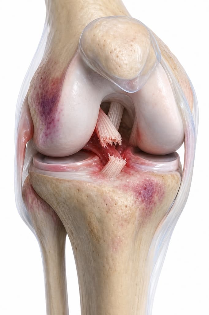
Diagram showing an anterior cruciate ligament tear inside the center of the knee joint.
Torn ACL Fibres

Mid-substance discontinuity of the stabilizing ligament.

Posterior Cruciate Ligament (PCL)

Remains intact behind the ACL, stabilizing posterior drawer.

Location of the Anterior Cruciate Ligament (ACL) and common mid-substance tear site.. (Pulsing letters show planned interactive annotation markers)
Torn ACL FibresPathology

Mid-substance discontinuity of the stabilizing ligament.

Posterior Cruciate Ligament (PCL)

Remains intact behind the ACL, stabilizing posterior drawer.

Anatomy Comparison

Healthy vs Affected Joint Comparison

Understanding structural differences caused by acl injury.
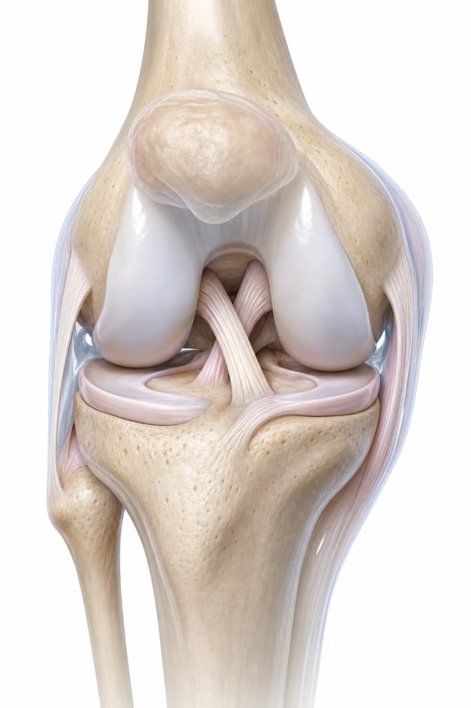
Normal Anatomy
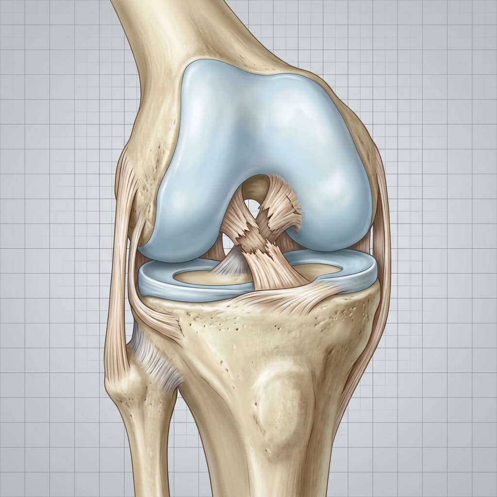
Normal AnatomyNormal ACLClinical Reference
Pathological / Affected Anatomy (Ligament Disruption)
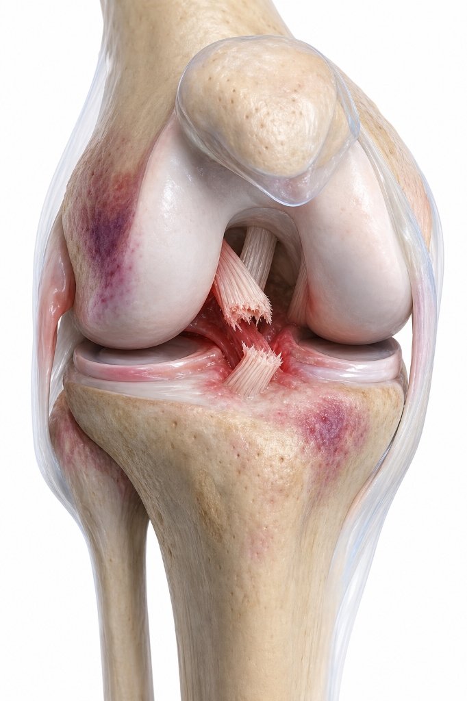
Pathological / Affected AnatomyTorn ACLClinical Reference
Ligament Integrity

Normal Anatomy

Taut, continuous Anterior Cruciate Ligament stabilizing the center of the joint

Pathological / Affected Anatomy

Disrupted, torn, or wavy ligament fibers lacking structural continuity

Joint Stability

Normal Anatomy

Tibia remains aligned under load (no abnormal forward sliding)

Pathological / Affected Anatomy

Excessive forward translation of the tibia (joint laxity/giving way)

Secondary Structures

Normal Anatomy

Intact surrounding bones and meniscus cartilage

Pathological / Affected Anatomy

Risk of bone bruising (contusion) or secondary meniscal tears from instability

Annotated Imaging Example

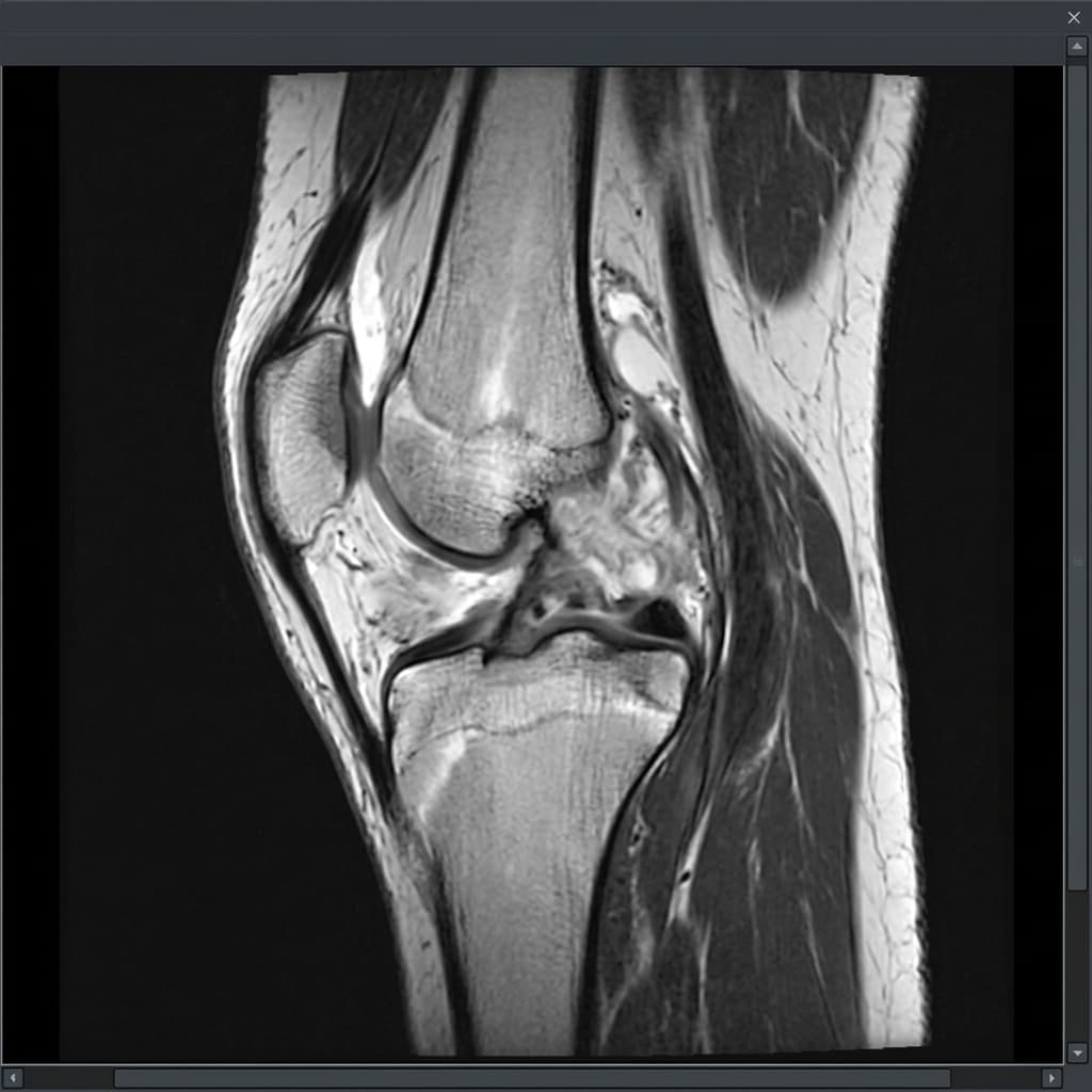
LKC Imaging Viewer
MRI · Sagittal T2-weighted
Sagittal MRI scan of the knee showing disruption of the anterior cruciate ligament with associated bone bruising. Annotations identify key structural changes.
Annotations visibleEducational use only
Sagittal T2 MRI demonstrating complete ACL disruption. Educational example only — not for diagnostic interpretation.
Annotation Reference
  1. 1ACL — Absent/Disrupted Signal
  2. 2Bone Bruising (Lateral Femoral Condyle)
  3. 3PCL — Intact
Educational Imaging DisclaimerThis imaging example is provided solely for educational purposes. It is not a diagnostic image and does not represent your own anatomy or pathology. All patients require individual clinical examination, history taking, and appropriate investigations to reach a diagnosis. The findings shown may not be present in your case.

Anatomy Comparison — Intact versus Torn ACL

Anatomy Comparison

Intact ACL vs Torn ACL

Educational comparison of normal ligament integrity and disrupted anatomy.
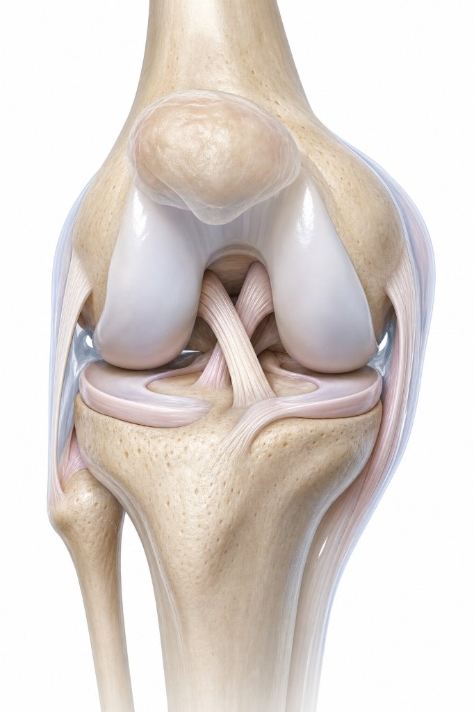
Intact ACL — Normal AnatomyNormal anatomy illustration
Torn ACL — Disrupted AnatomyAffected anatomy illustration
Both panels are educational illustrations. They do not represent any individual patient's anatomy. ACL injury diagnosis requires clinical examination and MRI.

Common symptoms

Symptoms vary, and similar symptoms can occur with other knee conditions. Commonly reported signs include:

A loud 'pop' or popping sensation at the time of injury
Severe pain and rapid joint swelling within a few hours
A feeling of instability, shift, or the knee 'giving way'
Difficulty bearing weight on the leg or walking normally

Causes and associated factors

The development or onset of this condition can be associated with several mechanical, biological, and lifestyle factors, including:

  • Sudden change of direction, twisting, or cutting movements
  • Landing awkwardly from a jump or stopping suddenly while running
  • Direct trauma to the outside of the knee (e.g., contact sports tackle)

Clinical assessment and diagnosis

A comprehensive clinical assessment is performed to understand the root cause of your symptoms and outline appropriate management options.

  • Discussion of symptoms, onset, and injury mechanism (e.g. non-contact pivot)
  • Physical examination checking joint swelling, tenderness, and ligament laxity
  • Laxity tests including Lachman's, Pivot Shift, and Anterior Drawer tests
  • Evaluation of patient goals, physical demands, and activity expectations
A diagnosis should not be made from website information alone.

Investigations

Imaging and other investigations may be recommended depending on clinical presentation. Common assessments include:

MRI scan to assess the integrity of the ACL fibers and check for associated meniscus, cartilage, or bone bruising injuries

Indicated where clinically appropriate following assessment.

Diagnostics & Imaging >

Standard X-rays to rule out fractures or bone avulsions

Indicated where clinically appropriate following assessment.

Diagnostics & Imaging >
LKC Imaging Console v1.0
Status: clinically approved
Sagittal MRI slice showing absence of the normal dark, taut diagonal ACL band, replaced by amorphous high signal.
Disorganized ACL Ligament notch

Wavy, disconnected fibers and fluid accumulation in the femoral notch.

Bone Contusion (Tibial)

Bone bruising from translation during the initial pivot injury.

Sagittal MRI scan showing a full-thickness rupture of the Anterior Cruciate Ligament (ACL).. (Weight-bearing views are standard diagnostic protocols)
Disorganized ACL Ligament notchPathology

Wavy, disconnected fibers and fluid accumulation in the femoral notch.

Bone Contusion (Tibial)Pathology

Bone bruising from translation during the initial pivot injury.

Treatment options

We offer structured, staged treatment pathways, focusing on conservative and non-surgical approaches. Suitability depends on individual assessment.

A

RICE protocol

Rest, Ice, Compression, Elevation) and initial swelling contro

Explore Treatment >
B

Specialist physical rehabilitation focused on hamstring, quadriceps, and core strength

Indicated where clinically appropriate.

Explore Treatment >
C

Proprioception and balance training to restore joint position sense

Indicated where clinically appropriate.

Explore Treatment >
D

Knee bracing in the early recovery or return-to-sport phase

Indicated where clinically appropriate.

Explore Treatment >
E

Surgical ACL reconstruction using a tendon graft for patients with persistent instability

Indicated where clinically appropriate.

Explore Treatment >

When might surgery be considered?

Surgical intervention may be discussed when non-operative treatments do not provide sufficient relief, symptoms significantly restrict daily function or quality of life, and clinical and imaging findings correlate.

ACL Reconstruction using a hamstring tendon graft

Consultant orthopaedic procedure.

ACL Reconstruction using a patellar or quadriceps tendon graft

Consultant orthopaedic procedure.

Choosing the right treatment

Decisions depend on a detailed diagnosis, symptom severity, patient goals, activity expectations, and general health history. We support shared decision-making, ensuring you fully understand the expected benefits, risks, and alternatives of each pathway.

Frequently Asked Questions

No. Reconstruction is not mandatory. Sedentary individuals or those who do not play pivoting sports can often achieve stable, pain-free knees through high-quality physical rehabilitation. Surgery is recommended for patients experiencing persistent instability or wishing to return to pivoting sports.
Recovery is a gradual, phased process. It typically takes 9 to 12 months of structured physical therapy to safely return to contact or pivoting sports, though daily light activities can be resumed much sooner.
Immediately after the injury, pain, swelling, and muscle inhibition may make walking difficult. Once the acute symptoms subside (often in 1-2 weeks), most patients can walk normally, though the knee may still feel unstable during twisting movements.
Initial exercises focus on restoring full range of motion and reducing swelling. Progressive strengthening of the hamstrings, quadriceps, and glutes is followed by balance and agility training.
It is diagnosed through a combination of clinical physical assessment (including the Lachman's test) and confirmed by an MRI scan.
Yes, bringing any previous imaging discs and reports to your consultation is highly recommended to assist your consultant in planning your care.
Clinically Reviewed
Mr Ricardo J Pacheco (GMC 4145976)Consultant Trauma & Orthopaedic Surgeon
Last reviewed: 29 July 2026
References
  1. North Tees and Hartlepool NHS Foundation Trust — Anterior Cruciate Ligament (ACL) Injury
  2. Royal Berkshire NHS Foundation Trust — ACL injury: non-operative management
  3. Guideline on anterior cruciate ligament injury - PMC - NIH (pmc.ncbi.nlm.nih.gov)
  4. Best Practice for Management of Anterior Cruciate Ligament (ACL) ... (boa.ac.uk)
  5. Management of Anterior Cruciate Ligament Injuries (aaos.org)
  6. Evidence-based recommendations for the management of ... (pmc.ncbi.nlm.nih.gov)

Would you like an individual knee assessment?

A consultation can help clarify the cause of your symptoms and discuss appropriate treatment options.

Face-to-face appointments are arranged through the reception team at the chosen clinic. Video consultations use Google Calendar where available.

Continue Your Knee Journey

Next Step: Explore Treatment Options

Treatment pathways range from physiotherapy and bracing to injections or surgical procedures, tailored to your symptoms, diagnosis, and physical goals.

Explore Treatments

Get notified about new content on ACL Injury

We'll only email you about updates on this specific topic — unsubscribe anytime.

Medical Disclaimer

This website provides educational and general information about knee symptoms, conditions, and treatments. The content is for informational purposes only and does not constitute medical advice, diagnosis, or a treatment plan. It should not be used as a substitute for a professional consultation, examination, or clinical decision-making by a qualified orthopaedic specialist.

If you are experiencing symptoms or are concerned about a knee problem, please arrange a clinical consultation. If you have urgent symptoms, severe pain, or are unable to put weight on your leg, please read our urgent advice guidance or contact emergency services immediately (dial 999 or NHS 111).

Contact us on WhatsApp — for administrative enquiries only. Please do not send confidential medical information.