
Meniscus Structure
Normal Anatomy
Smooth, continuous C-shaped cartilage discs (medial and lateral)
Pathological / Affected Anatomy
Frayed, torn, or displaced cartilage segment (e.g. bucket-handle or flap tear)
A meniscal tear involves damage to the C-shaped shock-absorbing cartilage discs situated inside the knee joint. This page explains the role of the meniscus, common symptoms, assessment, and treatment pathways.
The medial and lateral menisci are vital C-shaped cartilage discs that act as shock absorbers, protecting the joint surfaces and distributing weight. Meniscal tears can occur from acute twisting injuries (common in sports) or as part of gradual degenerative changes in older joints. Wording is cautious: not all tears cause symptoms, and not all require surgery. Suitability for repair or conservative care depends on tear configuration, location, and clinical findings.
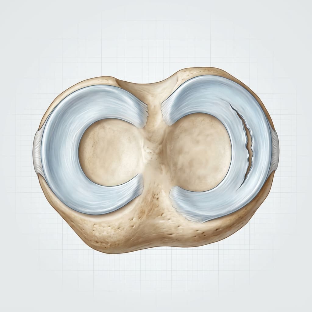
C-shaped inner cartilage shock absorber, frequently torn.
More circular outer cartilage shock absorber.
Typical tear occurring in the posterior horn of the medial meniscus.
C-shaped inner cartilage shock absorber, frequently torn.
More circular outer cartilage shock absorber.
Typical tear occurring in the posterior horn of the medial meniscus.
Understanding structural differences caused by meniscal tear.
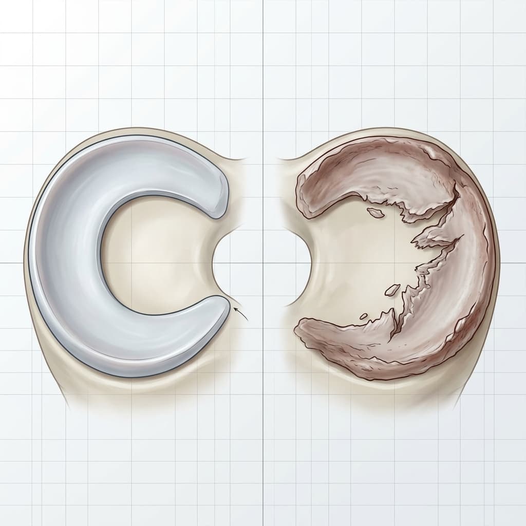 Comparison DiagramClinical Reference
Normal Anatomy
Smooth, continuous C-shaped cartilage discs (medial and lateral)
Pathological / Affected Anatomy
Frayed, torn, or displaced cartilage segment (e.g. bucket-handle or flap tear)
Normal Anatomy
Distributes weight-bearing forces evenly across the joint
Pathological / Affected Anatomy
Concentrated pressure on articular cartilage, potentially leading to pain and wear
Normal Anatomy
Glides smoothly during knee flexion and extension
Pathological / Affected Anatomy
Catching, clicking, or locking of the joint if a torn segment displaces
| Anatomical Feature | Normal Anatomy | Pathological / Affected Anatomy |
|---|---|---|
| Meniscus Structure | Smooth, continuous C-shaped cartilage discs (medial and lateral) | Frayed, torn, or displaced cartilage segment (e.g. bucket-handle or flap tear) |
| Shock Absorption | Distributes weight-bearing forces evenly across the joint | Concentrated pressure on articular cartilage, potentially leading to pain and wear |
| Mechanical Clearance | Glides smoothly during knee flexion and extension | Catching, clicking, or locking of the joint if a torn segment displaces |
Symptoms vary, and similar symptoms can occur with other knee conditions. Commonly reported signs include:
The development or onset of this condition can be associated with several mechanical, biological, and lifestyle factors, including:
A comprehensive clinical assessment is performed to understand the root cause of your symptoms and outline appropriate management options.
Imaging and other investigations may be recommended depending on clinical presentation. Common assessments include:
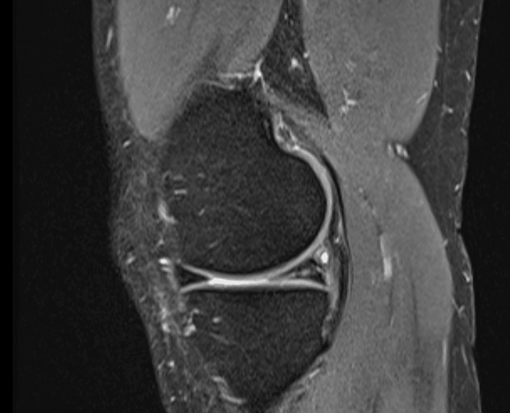
the gold standard for visualizing meniscus tear size and position
to check for joint degeneration or loose bony fragments
White linear signal extending to the joint surface indicating a tear.
White linear signal extending to the joint surface indicating a tear.
We offer structured, staged treatment pathways, focusing on conservative and non-surgical approaches. Suitability depends on individual assessment.
Indicated where clinically appropriate.
Explore Treatment >Indicated where clinically appropriate.
Explore Treatment >Surgical intervention may be discussed when non-operative treatments do not provide sufficient relief, symptoms significantly restrict daily function or quality of life, and clinical and imaging findings correlate.
stitching the tear back together for vascularized tears
trimming away unstable torn cartilage edges
Decisions depend on a detailed diagnosis, symptom severity, patient goals, activity expectations, and general health history. We support shared decision-making, ensuring you fully understand the expected benefits, risks, and alternatives of each pathway.
A consultation can help clarify the cause of your symptoms and discuss appropriate treatment options.
Face-to-face appointments are arranged through the reception team at the chosen clinic. Video consultations use Google Calendar where available.
Treatment pathways range from physiotherapy and bracing to injections or surgical procedures, tailored to your symptoms, diagnosis, and physical goals.
Get notified about new content on Meniscal Tear
We'll only email you about updates on this specific topic — unsubscribe anytime.
This website provides educational and general information about knee symptoms, conditions, and treatments. The content is for informational purposes only and does not constitute medical advice, diagnosis, or a treatment plan. It should not be used as a substitute for a professional consultation, examination, or clinical decision-making by a qualified orthopaedic specialist.
If you are experiencing symptoms or are concerned about a knee problem, please arrange a clinical consultation. If you have urgent symptoms, severe pain, or are unable to put weight on your leg, please read our urgent advice guidance or contact emergency services immediately (dial 999 or NHS 111).