
Key Takeaways
- Articular cartilage is a highly specialized, friction-free tissue that cushions joint bones.
- Early cartilage wear occurs silently because the tissue lacks blood vessels and nerves.
- Osteoarthritis is a dynamic process involving cartilage loss, bone spur development, and inflammation.
- Weight-bearing X-rays and detailed MRIs are used to accurately diagnose and grade joint wear.
- Joint preservation strategies focus on mechanical unloading and protecting remaining cartilage.
Understanding Your Knee Health: The Biology of Articular Cartilage
Within your knee joint, the ends of the femur (thighbone) and tibia (shinbone) are capped by a highly specialized structure called articular (hyaline) cartilage. Only a few millimeters thick, this tissue is incredibly resilient, elastic, and smooth—possessing a coefficient of friction that is slipperier than ice sliding on ice. Articular cartilage is composed of specialized cells called chondrocytes embedded within a dense matrix of collagen fibers (predominantly Type II) and water-binding proteins (proteoglycans, such as aggrecan). Its primary function is to distribute weight-bearing loads evenly across the joint and prevent bone-on-bone contact during movement.
Hyaline cartilage is structured in four distinct zones: the superficial zone, middle (transitional) zone, deep zone, and the calcified zone (separated by the tide mark). The superficial zone contains tightly packed collagen fibers oriented parallel to the joint surface, providing resistance to shear stress. The middle zone transition contains random collagen fibers and provides resistance to compression forces, while the deep zone features vertical columns of chondrocytes and thick collagen fibers anchored to the underlying bone. Because of this specialized structure, cartilage can absorb repetitive impacts for decades, but its lack of blood vessels, nerves, and lymphatic drainage means it has a very limited capacity to heal once damaged.
"Cartilage contains no blood vessels or nerves, meaning early wear and tear does not cause pain. Pain only arises when wear reaches the bone beneath, exposing sensitive nerve endings."
How Joint Wear Occurs: The Osteoarthritis Process
Because articular cartilage has no direct blood supply, it has a very limited capacity to salvage itself once damaged. Instead, it relies on joint movement to circulate synovial fluid, which delivers nutrients and removes cellular waste. When mechanical wear or injury disrupts this system, cartilage begins a process of degeneration.
Initially, the smooth surface of the cartilage develops microscopic cracks and rough patches, a stage known as fibrillation. As the wear progresses, the cartilage layer grows thinner, and small pieces may break away into the joint space. In response to this loss of cushioning, the underlying bone experiences increased stress and reacts by thickening (subchondral sclerosis) and growing bony projections at the joint edges (osteophytes, or bone spurs). These spurs are the body's attempt to distribute load over a larger surface area, but they can cause stiffness and pinch surrounding tissues, leading to inflammation of the synovial membrane.
Signs and Symptoms of Cartilage Wear
While early degeneration is silent, progressive cartilage loss eventually produces distinct clinical symptoms that impact daily life:
- •Morning Stiffness: The joint feels stiff and hard to bend upon waking, usually improving within 30 minutes as movement circulates synovial fluid.
- •Crepitus: A grinding, popping, or crunching sensation felt or heard when bending the knee, caused by rough cartilage surfaces rubbing together.
- •Activity-Induced Ache: A dull, deep ache inside the knee that worsens after prolonged walking, standing, or climbing stairs, and improves with rest.
- •Joint Swelling: Often called 'water on the knee,' this occurs when cartilage fragments irritate the synovial lining, prompting it to produce excess fluid.
Diagnosing and Grading Joint Wear: The Clinical Standard
To determine the extent of joint wear, knee specialists utilize specific diagnostic imaging tools. Standing, weight-bearing X-rays are the primary tool used to assess joint health. Because cartilage is transparent on X-rays, doctors look at the gap between the bones (the joint space). A narrowed joint space indicates cartilage thinning, while bone spurs and subchondral bone changes confirm arthritis.
- •In clinical practice, osteoarthritis is commonly graded using the Kellgren-Lawrence (KL) scale:
- •Grade 1 (Doubtful): Minimal joint space narrowing, possible tiny bone spurs.
- •Grade 2 (Mild): Definite bone spurs, minimal joint space narrowing.
- •Grade 3 (Moderate): Moderate joint space narrowing, multiple spurs, possible bone deformity.
- •Grade 4 (Severe): Severe joint space narrowing (bone-on-bone contact), large spurs, marked bone deformity.
For localized cartilage injuries (such as sports injuries), a high-resolution MRI is used to visualize focal defects, check the health of the underlying bone, and evaluate for surrounding pathology like meniscus tears or joint capsule thickening.

Protecting Your Remaining Cartilage: Preservation Strategies
While lost hyaline cartilage cannot be naturally regenerated, you can take active steps to preserve and protect your remaining joint structures:
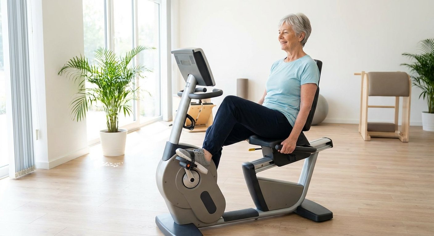
- •Maintain Low-Impact Movement: Regular cycling or swimming keeps the joint mobile and circulating synovial fluid without the impact forces of running.
- •Build Surrounding Strength: Strengthening your quadriceps and hip muscles provides a muscular shield that absorbs ground forces.
- •Manage Mechanical Alignment: Using offloading knee braces or orthotic shoe inserts can shift pressure away from the most worn areas of the joint.
- •Explore Biologics and Lubricants: Treatments like PRP or hyaluronic acid injections help manage joint chemistry and reduce the inflammatory environment that accelerates cartilage breakdown.