Lincolnshire Knee Clinic
Conditions

Patellar Instability and Kneecap Dislocation

Patellar instability occurs when the kneecap moves abnormally or dislocates from its usual position. This page explains common symptoms, assessment and treatment options.

On this page

What is Patellar Instability and Kneecap Dislocation?

The kneecap (patella) normally moves within a groove at the front of the thigh bone (femur), known as the trochlear groove. Patellar instability is a clinical term used when the kneecap moves abnormally within or outside this groove. This may involve partial displacement (subluxation) or complete dislocation. Symptoms vary considerably between individuals, and the degree of instability does not always reflect how much discomfort a patient experiences. Instability may follow a specific injury or may recur over time without a single clear cause.
This information is for general patient education and does not replace an individual clinical assessment.

Anatomical Overview

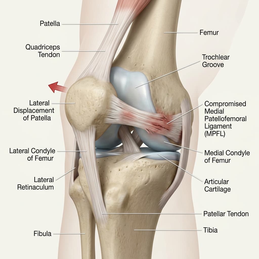
Anterior diagram showing a kneecap displacing laterally outside the boundaries of the trochlear groove.
Lateral patellar dislocation showing rupture of the stabilizing medial patellofemoral ligament (MPFL)..
Anatomy Comparison

Healthy vs Affected Joint Comparison

Understanding structural differences caused by patellar instability and kneecap dislocation.
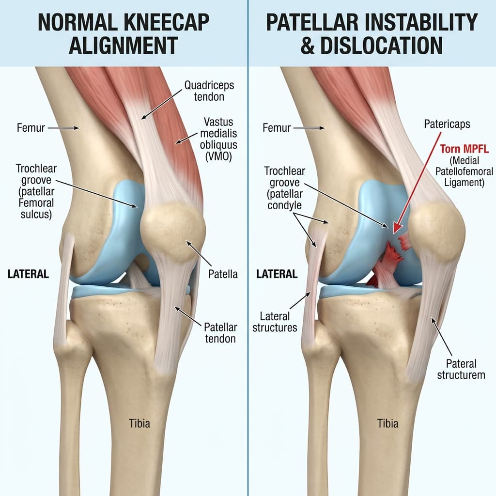
Healthy vs Affected Joint ComparisonComparison DiagramClinical Reference
Kneecap Position

Normal Anatomy

Kneecap sits securely inside the trochlear groove

Pathological / Affected Anatomy

Kneecap displaces to the outside (lateral subluxation/dislocation)

Stabilizing Ligaments

Normal Anatomy

Intact medial patellofemoral ligament (MPFL) preventing lateral slip

Pathological / Affected Anatomy

Stretched or torn MPFL allowing the patella to escape

Annotated Imaging Example

LKC Imaging Viewer
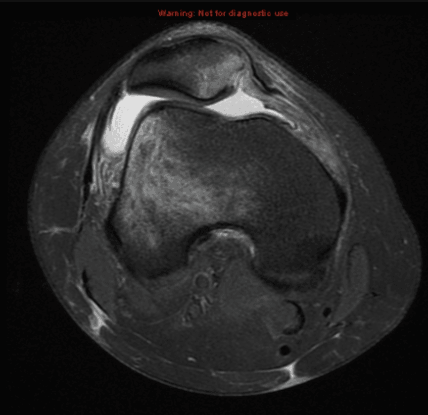
MRI · Axial MRI
Axial MRI of the patellofemoral joint showing patellar tracking and trochlear groove anatomy, annotated for educational use.
Annotations visibleEducational use only
Axial MRI demonstrating patellar position within the trochlear groove. Educational example only — not for diagnostic use.
Annotation Reference
  1. 1Patellar Tilt / Position
  2. 2Trochlear Groove Contour
  3. 3Medial Patellofemoral Ligament (MPFL) Region
Educational Imaging DisclaimerThis imaging example is provided solely for educational purposes. It is not a diagnostic image and does not represent your own anatomy or pathology. All patients require individual clinical examination, history taking, and appropriate investigations to reach a diagnosis. The findings shown may not be present in your case.

Common symptoms

Symptoms vary, and similar symptoms can occur with other knee conditions. Commonly reported signs include:

Sudden kneecap displacement or a sensation that the kneecap has moved
Pain around or behind the kneecap
Swelling following an episode of instability
Apprehension or fear that the kneecap may dislocate when bending or twisting
Recurrent giving way of the knee
Difficulty twisting, changing direction, or pivoting
Pain on stairs or during squatting movements
Reduced confidence in the knee during activity

Causes and associated factors

The development or onset of this condition can be associated with several mechanical, biological, and lifestyle factors, including:

  • Traumatic injury causing the kneecap to be pushed out of its groove
  • Previous kneecap dislocation (which itself increases the risk of recurrence)
  • Injury to the medial patellofemoral ligament (MPFL), which helps hold the kneecap in place
  • Trochlear shape (shallow or absent groove may allow the kneecap to displace more easily)
  • Patellar height (a high-riding kneecap may engage the groove later during bending)
  • Limb or rotational alignment factors that alter the direction of force through the kneecap
  • Muscle control and strength imbalances around the hip and thigh
  • Ligamentous laxity or generalised joint hypermobility
  • Sporting activity involving sudden pivoting, twisting, or change of direction

Clinical assessment and diagnosis

A comprehensive clinical assessment is performed to understand the root cause of your symptoms and outline appropriate management options.

  • Discussion of the mechanism of injury and how instability episodes occur
  • Clarification of whether this is a first-time or recurrent dislocation or subluxation
  • Assessment of swelling and any joint effusion
  • Review of any previous episodes, operations, or physiotherapy
  • Examination of kneecap tracking, position, and glide
  • Assessment for apprehension when the kneecap is displaced laterally
  • Review of limb alignment and rotational profile
  • Assessment of ligament stability, range of motion, and muscle strength
  • Discussion of patient goals, sporting demands, and activity requirements
A diagnosis should not be made from website information alone.

Investigations

Imaging and other investigations may be recommended depending on clinical presentation. Common assessments include:

X-rays: to assess kneecap position, trochlear shape, and bony anatomy

Indicated where clinically appropriate following assessment.

Diagnostics & Imaging >

MRI: to assess the MPFL, articular cartilage, and any soft-tissue injury following dislocation

Indicated where clinically appropriate following assessment.

Diagnostics & Imaging >

CT: occasionally used in selected cases to assess alignment, rotational anatomy, or trochlear morphology

Indicated where clinically appropriate following assessment.

Diagnostics & Imaging >
LKC Imaging Console v1.0
Status: clinically approved
Axial MRI of the patellofemoral joint showing patellar tracking and trochlear groove anatomy.
Patellar Tilt / Position

Position of the patella relative to the trochlear groove.

Trochlear Groove Contour

Cartilage-lined groove the patella tracks within.

Medial Patellofemoral Ligament (MPFL) Region

Medial soft tissue restraint adjacent to the medial border of the patella.

Axial MRI demonstrating patellar position within the trochlear groove. Educational example only — not for diagnostic use.. (Weight-bearing views are standard diagnostic protocols)
Patellar Tilt / PositionPathology

Position of the patella relative to the trochlear groove.

Trochlear Groove Contour

Cartilage-lined groove the patella tracks within.

Medial Patellofemoral Ligament (MPFL) Region

Medial soft tissue restraint adjacent to the medial border of the patella.

Treatment options

We offer structured, staged treatment pathways, focusing on conservative and non-surgical approaches. Suitability depends on individual assessment.

A

Activity modification to reduce loading on the patellofemoral joint during the acute phase

Indicated where clinically appropriate.

Explore Treatment >
B

Physiotherapy and rehabilitation focusing on quadriceps, hip, and core strengthening

Indicated where clinically appropriate.

Explore Treatment >
C

Swelling and pain management in the early stages

Indicated where clinically appropriate.

Explore Treatment >
D

Knee bracing where appropriate to support the kneecap and improve confidence

Indicated where clinically appropriate.

Explore Treatment >
E

Progressive strengthening and movement control exercises

Indicated where clinically appropriate.

Explore Treatment >
F

Return-to-sport rehabilitation programme for athletes

Indicated where clinically appropriate.

Explore Treatment >
G

Surgery in selected cases of recurrent instability or where non-surgical management has not resolved symptoms

Indicated where clinically appropriate.

Explore Treatment >

When might surgery be considered?

Surgical intervention may be discussed when non-operative treatments do not provide sufficient relief, symptoms significantly restrict daily function or quality of life, and clinical and imaging findings correlate.

MPFL reconstruction: reconstructing the medial patellofemoral ligament to restore kneecap stability, commonly considered after recurrent dislocations

Consultant orthopaedic procedure.

Tibial tubercle osteotomy: a bony realignment procedure considered in selected cases where patellar height or alignment is a contributing factor

Consultant orthopaedic procedure.

Trochleoplasty: a procedure to deepen the trochlear groove, considered only in carefully selected cases where trochlear dysplasia is severe

Consultant orthopaedic procedure.

Treatment of associated cartilage injury: where a dislocation has caused damage to the articular cartilage surface

Consultant orthopaedic procedure.

Choosing the right treatment

Decisions depend on a detailed diagnosis, symptom severity, patient goals, activity expectations, and general health history. We support shared decision-making, ensuring you fully understand the expected benefits, risks, and alternatives of each pathway.

Frequently Asked Questions

Not every first-time dislocation requires surgery. Many patients recover well with physiotherapy and rehabilitation. Surgery may be considered in cases of recurrent instability or where specific anatomical factors are contributing. An individual assessment is needed to determine the most appropriate pathway.
A subluxation is a partial displacement of the kneecap from its groove, where it moves abnormally but may return to position on its own. A dislocation is a complete displacement, where the kneecap moves entirely out of the groove and typically requires manual reduction. Both can cause significant symptoms.
Not every patient requires an MRI. Imaging is determined by the clinical assessment and history. MRI may be particularly useful after a first dislocation to assess the MPFL, articular cartilage, and other soft-tissue structures. Your surgeon will advise based on your specific situation.
Yes. Physiotherapy plays an important role in patellar instability management, focusing on strengthening the muscles around the hip, quadriceps, and core to improve kneecap tracking and joint stability. Physiotherapy is often the first-line treatment for first-time dislocations.
Recurrent instability may occur because of anatomical factors (such as trochlear shape or patellar height), soft-tissue factors (such as MPFL laxity following initial injury), or muscle control issues. An individual assessment with imaging can help identify any contributing factors.
Surgery may be considered in cases of recurrent patellar instability that has not responded to adequate physiotherapy, or in first-time cases where specific anatomical risk factors are present. The surgical approach depends on the individual anatomy, symptoms, and patient goals and is discussed in a shared decision-making consultation.
Many patients with patellar instability are able to return to sport following appropriate treatment and rehabilitation. However, return to sport depends on the individual clinical situation, treatment pathway, rehabilitation progress, and the specific demands of the sport. No guarantee of return to any activity can be made without an individual assessment.
A GP referral is recommended for patients using private medical insurance, as most insurers require it to authorise cover. Self-paying patients can typically self-refer directly to the clinic. Please check your insurance policy prior to booking.
Clinically Reviewed
Mr Ricardo J Pacheco (GMC 4145976)Consultant Trauma & Orthopaedic Surgeon
Last reviewed: 29 July 2026
References
  1. University Hospitals Sussex NHS Foundation Trust — Dislocation of your patella (kneecap)
  2. Norfolk & Waveney Community MSK — Patella (Kneecap) Dislocation
  3. Updated treatment guidelines for patellar instability: “un menu à la ... (pmc.ncbi.nlm.nih.gov)
  4. [PDF] Patella Dislocation - Physiotherapy Management - CHW (resources.schn.health.nsw.gov.au)
  5. Dislocated kneecap - NHS (nhs.uk)
  6. Patellofemoral instability - Right Decisions - NHS Scotland (rightdecisions.scot.nhs.uk)

Would you like an individual knee assessment?

A consultation can help clarify the cause of your symptoms and discuss appropriate treatment options.

Face-to-face appointments are arranged through the reception team at the chosen clinic. Video consultations use Google Calendar where available.

Continue Your Knee Journey

Next Step: Explore Treatment Options

Treatment pathways range from physiotherapy and bracing to injections or surgical procedures, tailored to your symptoms, diagnosis, and physical goals.

Explore Treatments

Get notified about new content on Patellar Instability and Kneecap Dislocation

We'll only email you about updates on this specific topic — unsubscribe anytime.

Medical Disclaimer

This website provides educational and general information about knee symptoms, conditions, and treatments. The content is for informational purposes only and does not constitute medical advice, diagnosis, or a treatment plan. It should not be used as a substitute for a professional consultation, examination, or clinical decision-making by a qualified orthopaedic specialist.

If you are experiencing symptoms or are concerned about a knee problem, please arrange a clinical consultation. If you have urgent symptoms, severe pain, or are unable to put weight on your leg, please read our urgent advice guidance or contact emergency services immediately (dial 999 or NHS 111).

Contact us on WhatsApp — for administrative enquiries only. Please do not send confidential medical information.