Lincolnshire Knee Clinic
Conditions

Knee Tendinopathy

Knee tendinopathy is an overuse condition affecting the tendons. This page covers loading principles, assessment, and progressive rehabilitation.

On this page

What is Knee Tendinopathy?

Knee tendinopathy is an overuse condition affecting the tendons surrounding the joint (most commonly the patellar tendon below the kneecap). It involves micro-tearing and collagen degeneration in response to repetitive loading. Wording is cautious: there is no guaranteed recovery time, and progressive loading is the gold standard for rehabilitation. Rest alone is rarely curative.
This information is for general patient education and does not replace an individual clinical assessment.

Anatomical Overview

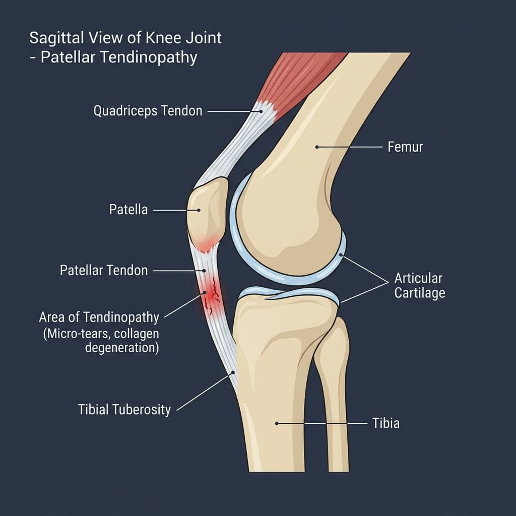
Diagram showing the patellar tendon connecting the kneecap to the shin bone with micro-tears highlighted.
Patellar tendinopathy affecting the proximal tendon attachment near the inferior pole of the patella..
Anatomy Comparison

Healthy vs Affected Joint Comparison

Understanding structural differences caused by knee tendinopathy.
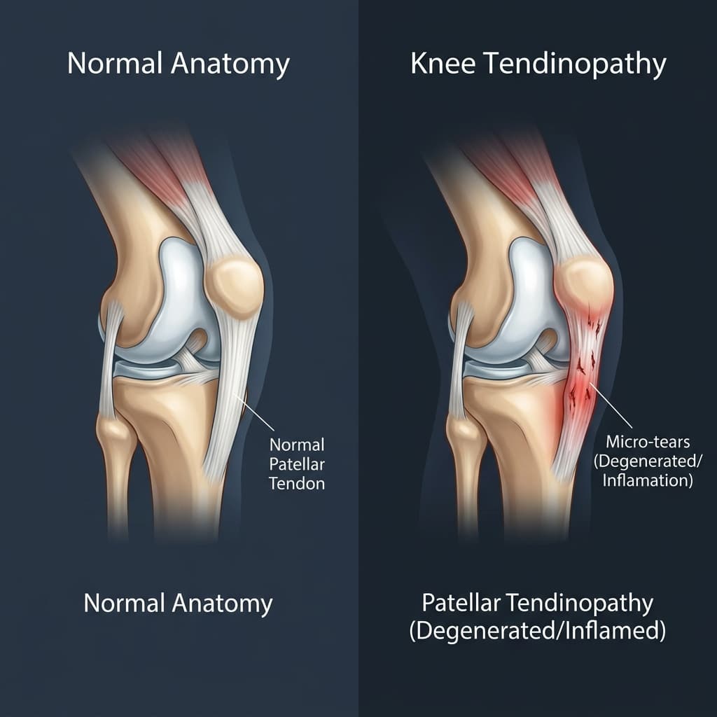
Healthy vs Affected Joint ComparisonComparison DiagramClinical Reference
Tendon Tissue

Normal Anatomy

Strong, organized collagen fibers without inflammation

Pathological / Affected Anatomy

Disorganized, thickened fibers, micro-tears, or local tenderness

Loading Response

Normal Anatomy

Pain-free force transfer during jumping, running, or stairs

Pathological / Affected Anatomy

Localized pain at the tendon insertion site under load

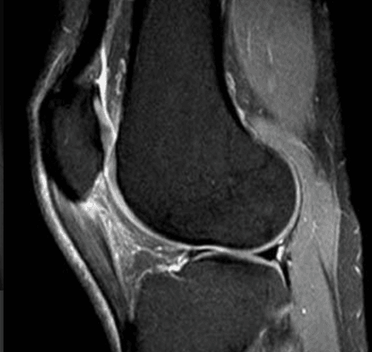
Annotated Imaging Example

LKC Imaging Viewer
MRI · Sagittal MRI
Sagittal MRI of the knee showing the joint and surrounding soft tissue, annotated for educational use.
Annotations visibleEducational use only
Sagittal MRI of the knee demonstrating patellar tendinopathy at the tendon's origin from the inferior pole of the patella. Educational example only.
Annotation Reference
  1. 1Patellar Tendinopathy
Educational Imaging DisclaimerThis imaging example is provided solely for educational purposes. It is not a diagnostic image and does not represent your own anatomy or pathology. All patients require individual clinical examination, history taking, and appropriate investigations to reach a diagnosis. The findings shown may not be present in your case.

Common symptoms

Symptoms vary, and similar symptoms can occur with other knee conditions. Commonly reported signs include:

Localized pain and tenderness over the tendon, worse at the start of load
Morning stiffness in the tendon that improves with light movement
Pain triggered by explosive actions like jumping, running, or squatting
Thickening or mild swelling of the tendon tissue

Causes and associated factors

The development or onset of this condition can be associated with several mechanical, biological, and lifestyle factors, including:

  • Repetitive overloading of the tendon without adequate rest
  • Sudden increases in training volume, intensity, or surface changes
  • Lower limb biomechanics or muscle tightness (e.g., tight quadriceps)
  • Inappropriate footwear or training surfaces

Clinical assessment and diagnosis

A comprehensive clinical assessment is performed to understand the root cause of your symptoms and outline appropriate management options.

  • Discussion of activity logs, training load, and symptom onset
  • Physical examination checking tenderness over the patellar or quadriceps tendon
  • Pain response testing during single-leg decline squats or hopping
  • Assessment of lower limb alignment and flexibility
A diagnosis should not be made from website information alone.

Investigations

Imaging and other investigations may be recommended depending on clinical presentation. Common assessments include:

Diagnostic ultrasound

preferred) to visualize tendon thickness, structure, and micro-tear

Diagnostics & Imaging >

MRI scan to evaluate the tendon in detail and rule out internal joint issues

Indicated where clinically appropriate following assessment.

Diagnostics & Imaging >
LKC Imaging Console v1.0
Status: clinically approved
Sagittal MRI of the knee showing the patellar tendon origin at the inferior pole of the patella.
Patellar Tendinopathy

Tendon origin at the inferior pole of the patella, where degenerative change occurs.

Sagittal MRI of the knee demonstrating patellar tendinopathy at the tendon's origin from the inferior pole of the patella.. (Weight-bearing views are standard diagnostic protocols)
Patellar TendinopathyPathology

Tendon origin at the inferior pole of the patella, where degenerative change occurs.

Treatment options

We offer structured, staged treatment pathways, focusing on conservative and non-surgical approaches. Suitability depends on individual assessment.

A

Progressive loading rehabilitation

eccentric or heavy slow resistance

Explore Treatment >
B

Activity modification and load management

Indicated where clinically appropriate.

Explore Treatment >
C

Extracorporeal Shockwave Therapy

ESWT) to stimulate tendon healin

Explore Treatment >
D

Targeted injection therapies in selected chronic cases

Indicated where clinically appropriate.

Explore Treatment >
E

Biomechanical adjustments and stretching

Indicated where clinically appropriate.

Explore Treatment >

When might surgery be considered?

Surgical intervention may be discussed when non-operative treatments do not provide sufficient relief, symptoms significantly restrict daily function or quality of life, and clinical and imaging findings correlate.

Tendon debridement or tenotomy in rare, recalcitrant cases failing rehab

Consultant orthopaedic procedure.

Choosing the right treatment

Decisions depend on a detailed diagnosis, symptom severity, patient goals, activity expectations, and general health history. We support shared decision-making, ensuring you fully understand the expected benefits, risks, and alternatives of each pathway.

Frequently Asked Questions

Tendonitis implies acute inflammation. Tendinopathy is chronic collagen degeneration from overload.
No. Complete rest makes the tendon weaker. Controlled, progressive loading is the key to recovery.
Healing can be slow due to limited tendon blood supply, often taking 3-6 months of rehabilitation.
A non-invasive treatment that uses acoustic waves to stimulate healing and blood flow in chronic tendons.
Yes, if load is modified. Exercise should be kept within a low, manageable pain threshold.
Recommended for private insurance; self-paying patients can self-refer.
Clinically Reviewed
Mr Ricardo J Pacheco (GMC 4145976)Consultant Trauma & Orthopaedic Surgeon
Last reviewed: 29 July 2026
References
  1. NHS Lanarkshire — Patellar Tendinopathy
  2. Musculoskeletal Matters (Dorset) — Knee pain: Patella tendinopathy
  3. Clinical Management of Patellar Tendinopathy - PMC - NIH (pmc.ncbi.nlm.nih.gov)
  4. Patellar tendinopathy advice leaflet (Jumper's knee) (ruh.nhs.uk)
  5. Patellar Tendinopathy (Jumper's Knee) - StatPearls - NCBI Bookshelf - NIH (ncbi.nlm.nih.gov)
  6. Dutch multidisciplinary guideline on anterior knee pain - PMC (pmc.ncbi.nlm.nih.gov)
  7. Rehab Practice Guidelines for: Patellar Tendinopathy - CDN (bpb-us-w2.wpmucdn.com)

Would you like an individual knee assessment?

A consultation can help clarify the cause of your symptoms and discuss appropriate treatment options.

Face-to-face appointments are arranged through the reception team at the chosen clinic. Video consultations use Google Calendar where available.

Continue Your Knee Journey

Next Step: Explore Treatment Options

Treatment pathways range from physiotherapy and bracing to injections or surgical procedures, tailored to your symptoms, diagnosis, and physical goals.

Explore Treatments

Get notified about new content on Knee Tendinopathy

We'll only email you about updates on this specific topic — unsubscribe anytime.

Medical Disclaimer

This website provides educational and general information about knee symptoms, conditions, and treatments. The content is for informational purposes only and does not constitute medical advice, diagnosis, or a treatment plan. It should not be used as a substitute for a professional consultation, examination, or clinical decision-making by a qualified orthopaedic specialist.

If you are experiencing symptoms or are concerned about a knee problem, please arrange a clinical consultation. If you have urgent symptoms, severe pain, or are unable to put weight on your leg, please read our urgent advice guidance or contact emergency services immediately (dial 999 or NHS 111).

Contact us on WhatsApp — for administrative enquiries only. Please do not send confidential medical information.